Jugular foramen
| Jugular foramen | |
|---|---|
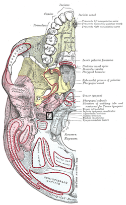 Base of skull. Inferior surface. (label for jugular foramen is at right, third from the bottom) | |
| Details | |
| Identifiers | |
| Latin | Foramen jugulare |
| TA | A02.1.00.054 |
| FMA | 56432 |
Anatomical terminology [edit on Wikidata] | |
The jugular foramen is a large foramen (aperture) in the base of the skull. It is located behind the carotid canal and is formed in front by the petrous portion of the temporal bone, and behind by the occipital bone; it is generally larger on the right than on the left side.
Contents
1 Contents
2 Clinical significance
3 Additional images
4 See also
5 References
6 External links
Contents
Cranial nerves IX (glossopharyngeal), X (vagus), and XI (accessory) and the internal jugular vein pass through the jugular foramen.
The jugular foramen may be subdivided into three compartments, each with their own contents.
- The anterior compartment transmits the inferior petrosal sinus
- The intermediate transmits the glossopharyngeal nerve, the vagus and accessory nerves (aka cranial nerves number IX, X, and XI respectively).
- The posterior transmits the sigmoid sinus (becoming the internal jugular vein) and some meningeal branches from the occipital and ascending pharyngeal arteries.
An alternative imaging based subclassification exists, delineated by the jugular spine which is a bony ridge partially separating the jugular foramen into two parts:
- The smaller, anteromedial, "pars nervosa" compartment contains CN IX, (tympanic nerve, a branch of CN IX), and receives the venous return from inferior petrosal sinus.
- The larger, posterolateral, "pars vascularis" compartment contains CN X, CN XI, Arnold's nerve (or the auricular branch of CN X involved in the Arnold's reflex, where external auditory meatus stimulation causes cough), jugular bulb, and posterior meningeal branch of ascending pharyngeal artery.
Clinical significance
Obstruction can result in "Vernet's syndrome".[1][2]
Additional images

Jugular foramen
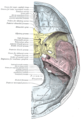
Base of the skull. Upper surface.


See also
- Occipitomastoid suture
References
This article incorporates text in the public domain from page 181 of the 20th edition of Gray's Anatomy (1918)
^ "Parapharyngeal Masses: Their Diagnosis and Management". Archived from the original on 2008-09-07..mw-parser-output cite.citation{font-style:inherit}.mw-parser-output q{quotes:"""""""'""'"}.mw-parser-output code.cs1-code{color:inherit;background:inherit;border:inherit;padding:inherit}.mw-parser-output .cs1-lock-free a{background:url("//upload.wikimedia.org/wikipedia/commons/thumb/6/65/Lock-green.svg/9px-Lock-green.svg.png")no-repeat;background-position:right .1em center}.mw-parser-output .cs1-lock-limited a,.mw-parser-output .cs1-lock-registration a{background:url("//upload.wikimedia.org/wikipedia/commons/thumb/d/d6/Lock-gray-alt-2.svg/9px-Lock-gray-alt-2.svg.png")no-repeat;background-position:right .1em center}.mw-parser-output .cs1-lock-subscription a{background:url("//upload.wikimedia.org/wikipedia/commons/thumb/a/aa/Lock-red-alt-2.svg/9px-Lock-red-alt-2.svg.png")no-repeat;background-position:right .1em center}.mw-parser-output .cs1-subscription,.mw-parser-output .cs1-registration{color:#555}.mw-parser-output .cs1-subscription span,.mw-parser-output .cs1-registration span{border-bottom:1px dotted;cursor:help}.mw-parser-output .cs1-hidden-error{display:none;font-size:100%}.mw-parser-output .cs1-visible-error{font-size:100%}.mw-parser-output .cs1-subscription,.mw-parser-output .cs1-registration,.mw-parser-output .cs1-format{font-size:95%}.mw-parser-output .cs1-kern-left,.mw-parser-output .cs1-kern-wl-left{padding-left:0.2em}.mw-parser-output .cs1-kern-right,.mw-parser-output .cs1-kern-wl-right{padding-right:0.2em}
^ Erol FS, Kaplan M, Kavakli A, Ozveren MF (June 2005). "Jugular foramen syndrome caused by choleastatoma". Clin Neurol Neurosurg. 107 (4): 342–6. doi:10.1016/j.clineuro.2004.08.006. PMID 15885397.
External links
| Wikimedia Commons has media related to Jugular foramen. |
Anatomy figure: 22:5b-12 at Human Anatomy Online, SUNY Downstate Medical Center
Anatomy figure: 22:4a-08 at Human Anatomy Online, SUNY Downstate Medical Center
cranialnerves at The Anatomy Lesson by Wesley Norman (Georgetown University) (IX, XI)
"Anatomy diagram: 34257.000-1". Roche Lexicon - illustrated navigator. Elsevier. Archived from the original on 2012-07-22.
- UCSD
Authority control |
|
|---|


